Renal Osteodystrophy
Understanding Bone Health in Chronic Kidney Disease
Overview
Renal osteodystrophy
When we think about kidney disease, we often focus on its impact on renal function and overall health. However, one lesser-known but significant complication of chronic kidney disease (CKD) is its effect on bone health. This condition, known as renal osteodystrophy, can have far-reaching consequences for patients with kidney problems.
Renal osteodystrophy is a complex bone disorder closely linked to chronic kidney disease (CKD). As kidney function declines, the body’s ability to regulate essential minerals and hormones becomes compromised, resulting in weakened and brittle bones. Early diagnosis and management can help improve the quality of life and reduce complications.
Why Is Bone Health Important?
Bones provide structure, protect vital organs, and store essential minerals like calcium and phosphorus. In individuals with CKD, imbalances in these minerals lead to poor bone health, increasing the risk of fractures, chronic pain, and mobility issues.
The Role of Healthy Kidneys in Bone Maintenance
Healthy kidneys play a key role in maintaining bone strength by:
- Regulating calcium and phosphorus levels.
- Producing calcitriol, the active form of vitamin D.
- Excreting excess minerals to maintain balance.
When kidney function is impaired, these processes are disrupted, leading to complications such as high phosphorus levels, low calcium levels, and an overproduction of parathyroid hormone (PTH), all of which weaken bones.
Types of Renal Osteodystrophy
Understanding the different forms of renal osteodystrophy helps in diagnosis and treatment:
Causes of Renal Osteodystrophy
Several factors contribute to the development of this condition, including:
- Imbalanced Minerals: Excess phosphorus and low calcium levels disrupt bone remodeling.
- High PTH Levels: Overactive parathyroid glands break down bone tissue to release calcium.
- Vitamin D Deficiency: Reduced calcitriol production leads to poor calcium absorption.
- Kidney Failure: Limits the body’s ability to filter and balance minerals effectively.
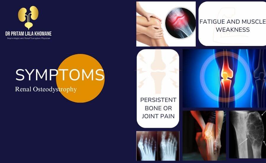
Symptoms to Watch For
While early stages may show no symptoms, advanced renal osteodystrophy can manifest as:
- Persistent bone or joint pain.
- Skeletal deformities, especially in children.
- Increased frequency of fractures.
- Fatigue and muscle weakness.
- Children with the condition may also experience growth delays or develop conditions like rickets.
Diagnosis of Renal Osteodystrophy
Timely diagnosis is critical to prevent severe complications. The following tests may be recommended:
- Blood Tests:
- Evaluate levels of calcium, phosphorus, PTH, and vitamin D.
- Assess kidney function using creatinine and eGFR levels.
- Bone Biopsy:
- Performed in rare cases to confirm diagnosis and assess bone structure.
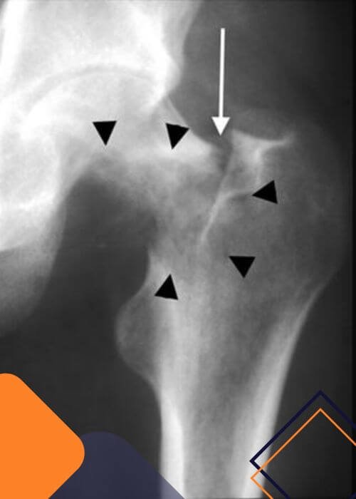
- Imaging Tests:
- X-rays, MRIs, or DEXA scans to evaluate bone density and detect deformities.
Treatment Options for Renal Osteodystrophy
Managing renal osteodystrophy focuses on restoring mineral balance, controlling CKD progression, and preventing bone damage.
Increase Calcium-Rich Foods: Leafy greens, fortified plant-based milks (within dietary restrictions).
Hydration: Adequate water intake helps flush excess phosphorus.
Vitamin D Supplements: Improve calcium absorption and reduce PTH levels.
Calcimimetics: Mimic calcium to reduce overactivity of parathyroid glands.
Recovery
Living with Renal Osteodystrophy
For Patients:
- Stay informed about your condition and follow your doctor’s advice.
- Focus on a kidney-friendly diet and stay active within your physical limits.
For Families:
- Support patients by ensuring adherence to treatment plans and dietary restrictions.
- Encourage open communication with healthcare providers.
consultation
When to Seek Medical Advice
Consult your doctor if you notice:
- Persistent or worsening bone pain.
- Difficulty performing daily activities due to muscle weakness.
- Unexplained fractures or deformities.
Meet the Nephrologist Who Can Help You with Renal Osteodystrophy
Dr. Pritam Lala Khomane specializes in renal osteodystrophy, offering personalized treatment plans and a multidisciplinary approach. With advanced techniques and patient-focused care, he ensures effective management and improved quality of life.
Acute Kidney Injury (AKI):
Frequently Asked Questions (FAQ)
What is renal osteodystrophy?
Renal osteodystrophy is a bone disease caused by chronic kidney disease (CKD). It results from imbalances in calcium, phosphorus, and vitamin D levels due to impaired kidney function, leading to weakened bones and an increased risk of fractures.
What causes renal osteodystrophy?
The condition is caused by:
- High levels of phosphorus in the blood.
- Low calcium levels due to poor absorption.
- Reduced production of vitamin D by the kidneys.
These disruptions are common in individuals with CKD.
What are the symptoms of renal osteodystrophy?
Common symptoms include:
- Bone pain.
- Increased risk of fractures.
- Growth delays in children.
- Skeletal deformities, such as rickets in children.
How is renal osteodystrophy diagnosed?
Diagnosis involves:
- Blood tests to measure calcium, phosphorus, vitamin D, and parathyroid hormone levels.
- Imaging tests like X-rays, DEXA scans, or MRIs to evaluate bone health.
- Bone biopsy (rarely) to analyze bone density and structure.
What are the treatment options for renal osteodystrophy?
Treatment focuses on managing mineral imbalances and includes:
- A low-phosphorus diet.
- Medications like phosphorus binders, calcium, and vitamin D supplements.
- Parathyroid surgery in severe cases.
Can renal osteodystrophy be prevented?
While it cannot be entirely prevented, progression can be slowed by:
- Managing CKD with proper medication and diet.
- Attending regular checkups to monitor mineral and hormone levels.
- Completing dialysis treatments as prescribed.
Why is it important to treat renal osteodystrophy?
Untreated renal osteodystrophy can lead to severe complications, such as fractures, skeletal deformities, and cardiovascular issues caused by mineral imbalances. Early diagnosis and treatment are essential to maintain bone and overall health.
Who should I consult for renal osteodystrophy treatment?
A nephrologist specializing in kidney diseases and related complications is the best healthcare professional to guide you in managing renal osteodystrophy.
If you have more questions, feel free to consult Dr. Pritam Lala Khomane, an expert nephrologist specializing in renal osteodystrophy care.
Gallery